

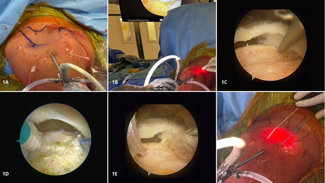
Figure 1A-F. Stepwise subacromial portal establishment and medial anchor placement during rotator cuff repair.
1A: Spinal needles define posterolateral and anterolateral portal positions that are used to facilitate viewing and suture management. 1B: A needle is used to localize percutaneous entry points for medial anchors while viewing from the lateral portal. 1C and 1D: A punch is advanced along the predefined trajectories to prepare the bone and insert the anteromedial and posteromedial anchors. 1E: Cannula placement between the medial anchors to facilitate suture management while viewing from the posterolateral portal. 1F: Illustration of the percutaneous placement of medial-row anchors.
1A: Spinal needles define posterolateral and anterolateral portal positions that are used to facilitate viewing and suture management. 1B: A needle is used to localize percutaneous entry points for medial anchors while viewing from the lateral portal. 1C and 1D: A punch is advanced along the predefined trajectories to prepare the bone and insert the anteromedial and posteromedial anchors. 1E: Cannula placement between the medial anchors to facilitate suture management while viewing from the posterolateral portal. 1F: Illustration of the percutaneous placement of medial-row anchors.
Courtesy of Ashraf Hantouly, MD, MSc, and Moin Khan, MD, MSc, FRCSC
Published 7/28/2026
|
Ashraf Hantouly, MD, MSc; Moin Khan, MD, MSc, FRCSC
At a Glance
- Prepare the footprint to promote healing while preserving bone integrity.
- Disorganized sutures create problems before knots are even tied.
- A broad surgical toolbox allows adaptation; a forced repair leads to failure.
Estimated read time: 4 minutes
Editor’s note: This article is part of “Orthopaedic Fundamentals,” a recurring AAOS Now series highlighting core principles widely embraced across the orthopaedic community. The series highlights ideas that can improve efficiency within and outside the OR, informing training surgeons as well as providing practical refreshers for experienced surgeons.
In this two-part installment on arthroscopic rotator cuff repair, part one reviewed preoperative planning, visualization, portal placement, and diagnostic arthroscopy. Part two focuses on tear assessment and the fundamental principles of repair.
Following diagnostic arthroscopy and establishment of adequate visualization, attention turns to the repair itself. Successful arthroscopic rotator cuff repair requires careful assessment of tear characteristics, thoughtful preparation of the biologic environment, and precise execution of fixation principles. Although repair techniques and implant technologies continue to evolve, the fundamental concepts underlying successful repair remain largely unchanged.
Tear assessment requires evaluation of pattern, mobility, and reducibility
Tear assessment should be performed only after achieving complete visualization. Tear pattern (crescent, U-shaped, L-shaped), tendon mobility, and reducibility must be evaluated dynamically using multiple viewing portals. The lateral and posterolateral portals typically provide the most informative perspective for rotator cuff repairs.
Adequate visualization, achieved through a thorough bursectomy and optimized portal placement, is a prerequisite before any repair is attempted.
Pearl: You cannot fix what you cannot see.
Prepare the rotator cuff footprint to optimize biology and healing
Optimizing the biological environment is critical to tendon healing. The rotator cuff footprint on the greater tuberosity should be prepared to promote bleeding and healing without excessive bone removal.
Gentle decortication using a shaver in forward mode is typically sufficient. Over-aggressive burring should be avoided, as it compromises bone quality and increases the risk of anchor pull-out. When using all-suture anchors, decortication is better avoided.
Pearl: Prepare the footprint to promote healing while preserving bone integrity.
---
Related Content
- Orthopaedic Fundamentals: Rotator cuff repair evaluation, visualization, and diagnostic arthroscopy (AAOS Now)
---
Apply technical fundamentals to ensure stable repair
While repair constructs may vary, core technical principles remain consistent.
Anchor placement
Anchor trajectory should be planned using a spinal needle. Entry points should be positioned as medially as possible (hugging the acromion), often just lateral to the humeral head articular margin, to optimize fixation strength and footprint coverage. Proper trajectory ensures effective medial-row fixation and facilitates subsequent lateral-row constructs when applicable (Figures 1B-F).
Arm positioning using a limb holder is critical to optimize visualization and anchor trajectory. For example, external rotation facilitates visualization and trajectory for the anteromedial anchor, whereas internal rotation improves access for the posteromedial anchor. Anchors are inserted through their respective anteromedial and posteromedial entry points while viewing from the lateral or posterolateral portal.
Pearl: Spinal needle and arm positioning are key tools to optimize anchor trajectory and facilitate accurate placement.
Suture passage
Sutures should be passed through robust tendon tissue while avoiding overly medial bites that increase repair tension. Symmetry in suture configuration is essential to achieve balanced load distribution. Efficient suture management is equally critical. Organized handling, use of color coding, and systematic sequencing reduce the risk of tangling and improve operative efficiency.
Pearl: Disorganized sutures create problems before knots are even tied.
Avoid these common mistakes
Common errors include over-tensioning the repair, inadequate medial fixation, and failure to address dog-ear deformities. When tissue quality is poor or mobility is limited, alternative strategies such as margin convergence, additional fixation points, or partial repair should be considered. Dog-ear deformities can be effectively addressed using luggage-tag sutures incorporated into lateral-row anchors. Anchor failure requires familiarity with implant-specific bailout strategies, including upsizing or altering fixation technique.
Pearl: A broad surgical toolbox allows adaptation; a forced repair leads to failure.
Final assessment and stability check
Before concluding the procedure, the surgeon must systematically evaluate the repair. This includes confirming adequate footprint coverage, symmetric suture tension, and a smooth tendon contour without gaps or dog-ears.
Dynamic assessment through gentle range-of-motion testing is essential to evaluate construct stability.
Pearl: Repair tension at rest predicts failure under load.
Conclusion
Rotator cuff repair succeeds through consistency, precision, and sound intraoperative judgment. Mastery of these fundamental principles — including tear assessment, biologic preparation, fixation strategy, and repair evaluation — provides a framework that remains applicable across evolving techniques and implant technologies. Although repair constructs will continue to advance, successful outcomes will remain rooted in careful execution of the fundamentals.
Ashraf Hantouly, MD, MSc, is a fellowship-trained orthopaedic surgeon with a specialized focus on shoulder and knee sports injuries.
Moin Khan, MD, MSc, FRCSC, is an associate professor of orthopaedic surgery at McMaster University, with a focus on sports medicine and shoulder surgery.